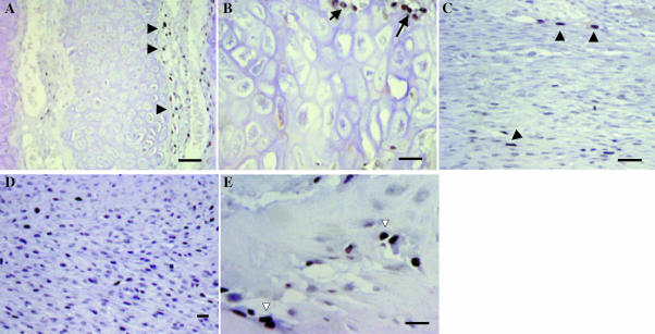
Fig. 6.
Proliferation in antler cartilage and at sites of intramembranous bone formation. PCNA staining (black). (A) Cartilage. Chondrocytes do not proliferate, but there are proliferating cells in the perivascular tissue that contains osteoblast and osteoclast precursors (arrowheads). (B) Higher power image of cartilage showing PCNA-positive cells close to vascular channels (arrows). (C) Few proliferating cells are detected in fibrous periosteum (arrowheads). (D) Proliferating cells are more abundant in cellular periosteum. (E) Bone. A proportion of osteoblasts are PCNA positive (open arrows). Bars: A, C, 100 µm; B, 50 µm; D, 25 µm; E, 20 µm.

