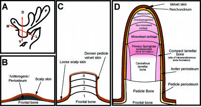
Fig. 2.
(A) Schematic diagram to show the three axes of the antler development: A-P, anterior-posterior axis; D-V, dorso-ventral axis; M-L, medio-lateral axis. (B,C) Schematic diagrams illustrating three stages of antler development. (B) Antlerogenic periosteum is present in the embryo and after birth as a localized thickening of the periosteum of the frontal bone. (C) Development of the pedicle occurs through four stages: 1, intramembranous ossification; 2, transitional ossification; 3, pedicle endochondral ossification; 4, antler endochondral ossification and velvet skin formation. (D) Longitudial section through a growing primary antler illustrating the main anatomical regions. Endochondral bone growth occurs at the distal tip while bone forms by intramembranous ossification around the antler shaft.

