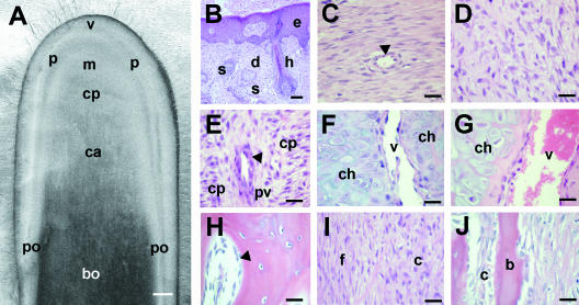
Fig. 9.
Histology of the regenerating antler during rapid longitudinal growth. (A) Longitudinal tissue section of antler tip to show macroscopic appearance of regions: v, velvet skin; p, perichondrium; m, mesenchyme; cp, chondroprogenitor region; c, cartilage; bo, bone; po, periosteum. Scale bar = 0.5 cm. (B–J) H&E-stained undecalcified paraffin sections of the tissue regions shown in A. (B) Velvet skin. e, epidermis; d, dermis; h, hair follicle; s, sebaceous gland. (C) Fibrous perichondrium. A blood vessel is marked by an arrowhead. (D) Mesenchymal ‘growth zone’. (E) Chondroprogenitor (cp) region. As in the early antler bud, cells start to become aligned in ‘columns.’ However, the vascular spaces are relatively small (arrowhead). (F) Non-mineralized cartilage. Recently differentiated chondrocytes (ch) are arranged in trabeculae separated by larger vascular channels (v). (G) Mineralized cartilage region. Chondrocytes and the vascular channels (v) increase in size in this region. (H) Spongy bone in the mid shaft of the antler that has formed by endochondral ossification. Osteoblasts are marked with an arrowhead. (I) Fibrous (f) and cellular (c) layers of the antler periosteum. (J) Intramembranous bone formation (b) takes place beneath the cellular periosteum (c). Scale bar (B–J), 100 µm.

