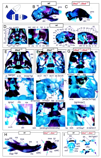
Fig. 11.
Skeletal analysis of the loss-of-function of the second-order paralogues, Dlx2−/−; Dlx5−/−, revealing a genetic interaction between the two. (A) Reference schema indicating the loss of two Dlx2 and two Dlx5 alleles in BA1. (B) Norma lateralis view of a P0 wild-type skull. (C) Norma lateralis view of a P0 Dlx2−/−; Dlx5−/− mutant skull. Note the rostrad displacement, apposition, and articulation of the truncated dentary (dnt) with the maxilla (mx) and not with the squamosal. (D) Wild-type P0 skulls highlighting, left to right, the palatal region (norma basalis; yellow line outlining the palatine and green line outlining the pterygoid), the ala temporalis and lamina obturans components of the alisphenoid, the ear region with the primary and secondary jaw articulations, and the middle ear without the dentary attached. (E, F) Norma basalis views of the Dlx5−/− (E) and Dlx2−/− (F) single mutants for comparison. (G) Skeletal staining of compound Dlx2−/−; Dlx5−/− neonate mutants emphasizing the drastic loss of BA structure. (H) Wild-type and Dlx2−/−; Dlx5−/− neonate PO dentaries. The mutant dentary is represented mainly by structures associated with the distal midline, such as the rostral process of Meckel's cartilage (rpMC). Mutant dentaries often possess an ectopic second incisor (e-lI). Green and black arrows point to miniscule cartilages associated with the proximal end of the truncated dentary. (I) Wild-type (left) and mutant (right) hyoid and thyroid cartilages. These caudal BA elements are also severely affected by the loss of both Dlx2 and Dlx5. The purple arrows are to highlight the asymmetric nature of the hyoids of these mutants. See text for detailed descriptions and list for abbreviations.

