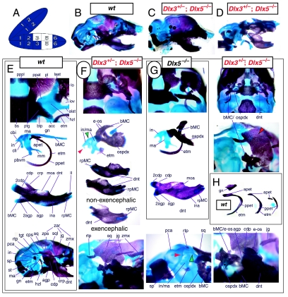
Fig. 15.
Skeletal analysis of the Dlx3+/−; Dlx5−/− mutant. (A) Reference schema indicating the loss of one Dlx3 allele and two Dlx5 alleles in BA1. (B) Norma lateralis view of a P0 wild-type skull. (C) Non-execephalic and(D) exencephalic norma lateralis views of P0 Dlx3+/−; Dlx5−/− mutant skulls. (E) Wild-type P0 skulls highlighting, top to bottom, norma basalis view of the ala temporalis and lamina obturans components of the alisphenoid, middle ear structures, and norma lateralis views of a wild-type mandible, and middle ear with the dentary attached. (F) P0 skulls of Dlx3+/−; Dlx5−/− mutants. The red arrowheads indicate the points of fusion between the head of the incus and the otic capsule, while the green arrows point to the fusion between the malleus and the crus brevis of the incus. The red and black arrowhead indicates the complete loss of the zygomatic process of the squamosal. (G) Dlx5−/− mutant middle ear structures and mandible for comparison. (H) Comparisons of wild-type and Dlx3+/−; Dlx5−/− ectotympanic development. See text for further descriptions and list for abbreviations.

