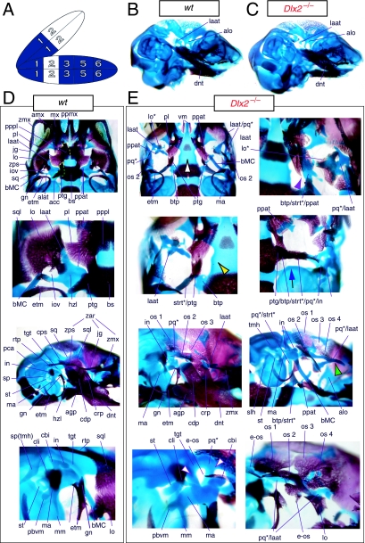
Fig. 4.
Skeletal analysis of Dlx2−/− mutants through differential staining of bone (alizarin red) and cartilage (alcian blue). (A) Reference schema indicating the loss of two Dlx2 alleles in BA1. (B) Wild-type E16.5 skull seen in oblique lateral view. (C) E16.5 skull of a Dlx2−/− mutant littermate showing a normal dentary (dnt) and the isolated tip of the lamina ascendens of the ala temporalis (laat). (D) Wild-type P0 skull highlighting, top to bottom, the palatal region (norma basalis; yellow line outlining the palatine and green line outlining the pterygoid), the ala temporalis and lamina obturans components of the alisphenoid, the ear region with the primary and secondary jaw articulations, and the middle ear. (E) P0 Dlx2−/− mutant skulls demonstrating alterations of mxBA1-derived structures. Purple arrowhead indicates an example exhibiting the loss of pterygoid bones. The green and black arrowhead points to the ectopic cartilage (pq*) running anterior from the region of the tegmen tympani and incorporating the lamina ascendens of the ala temporalis (laat). The purple and white arrowheads indicate the disassociation of the crus longus and crus brevis of the incus (cli and cbi), and the association of the crus longus with the tegman tympani (tgt), ectopic ‘strut’, and ectopic palatoquadrate (‘pq*’) cartilage. The blue and black arrow indicates the loss of the alicochlear commissure, while the yellow and black arrowhead indicates the ectopic lateral projection from the trabecular basal plate rostral to the basisphenoid body. See text for detailed descriptions and list for abbreviations.

