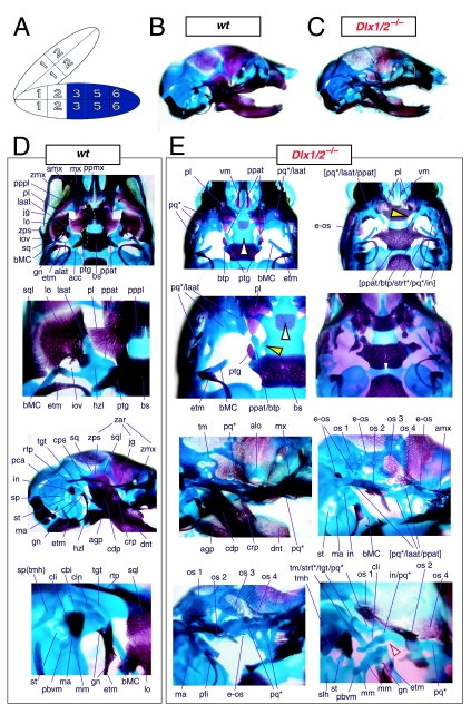
Fig. 6.
Skeletal analysis of compound Dlx1/2−/− mutants through differential staining of bone (alizarin red) and cartilage (alcian blue). (A) Reference schema indicating the loss of four Dlx alleles, two Dlx1 and two Dlx2, in BA1.(B) Norma lateralis view of a P0 wild-type skull. (C) Norma lateralis view of a P0 Dlx1/2−/− mutant skull. (D) Wild-type P0 skulls highlighting, top to bottom, the palatal region (norma basalis; yellow line outlining the palatine and green line outlining the pterygoid), the ala temporalis and lamina obturans components of the alisphenoid, norma lateralis view of the ear region with the primary and secondary jaw articulations and the middle ear. (E) P0 Dlx1/2−/− mutant skulls exhibiting alterations of mxBA1-derived structures. The yellow and black arrowheads point to cylindrical cartilages (ppat) running rostrad, parallel to the neurocranial base, toward the medial margins of the PQ*/lamina ascendens (pq*/laat) that have fused to the basitrabecular processes. Black and white arrowheads indicate the clefting of the palate. The red and white arrowhead highlights the loss of the crus brevis of the incus, and the fusion of the remainder of the incus to the ectopic strut (strt*) and palatoquadrate (pq*) structures. See text for detailed descriptions and list for abbreviations.

