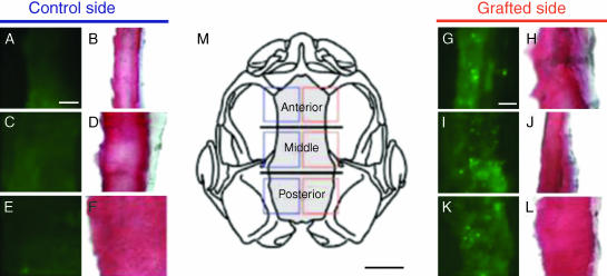
Fig. 6.
Neural crest derivation of the frontoparietal bone in Xenopus laevis, assessed by using fluorescein dextran. Fluorescent (A, C, E and G, I, K) and bright-field (B, D, F and H, J, L) images of cryosections through the frontoparietal bone in chimeric froglets, which earlier received labelled unilateral grafts of cranial neural crest. Frontal sections; anterior at top. Successive panels correspond to squares that are superimposed on the skull outlined in M (dorsal view; frontoparietal bone grey). Grafting labelled mandibular neural crest yields brightly labelled bone matrix in the rostral frontoparietal (G, green punctate clusters); labelling of bone matrix is confirmed by TriChrome staining in the following section (H, red). Similarly, intermediate (I, J) and caudal (K, L) portions of the bone are labelled by cells derived from hyoid and branchial crest stream grafts, respectively. Fluorescent marker is absent on the control (left) side of the skull (A–F), which did not receive labelled grafts. Horizontal lines in M demarcate three equal-sized zones in the frontoparietal bone, which were defined for the purposes of analysis. Scale bars: A–L, 25 mm; M, 1 mm. Reproduced with permission from Gross & Hanken (2005).

