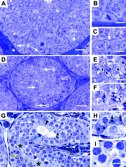
Fig. 1.
Histology of seminiferous epithelium in adult wild-type (A–C), 10-day-old wild-type (D–F) and adult hpg testis (G–I). (A) Adult wild-type seminiferous epithelium at stage IX of spermatogenic cycle showing nuclei of Sertoli cells (S), spermatogonium (SG), pachytene primary spermatocytes (P) and early elongating spermatids (SD). (B) Detail of a mature Sertoli cell nucleus with a tripartite nucleolus. (C) Highmagnification of A showing leptotene (L) and pachytene (P) primary spermatocytes between which is a thin curvilinear structure (arrow) representing inter-Sertoli cell junctional complex. (D) Ten-day wild-type testis showing basal and central Sertoli cell nuclei (S), spermatogonia (SG) and occasional spermatocytes (arrow). (E) sDetail of Sertoli cell nuclei showing ovoid and angular shapes with small nucleoli (arrowheads) and heterochromatin patches (arrows). (F) Most advanced germ cell type is occasional primary spermatocytes showing condensed chromatin (arrows). (G) hpg testis cord showing basal and central Sertoli cell nuclei (S), spermatogonia (SG) and central confluence of Sertoli cell cytoplasm (asterisks). Degenerative cellular debris (arrows) and hydropic-type germ cells are indicated (arrowhead). (H) Detail of Sertoli cell nuclei showing large and double-nucleoli (arrows). (I) Most advanced germ cells are zygotene primary spermatocytes with thickened chromatin profiles. Scale bars in A, D, G, = 20 µm; all others = 10 µm.

