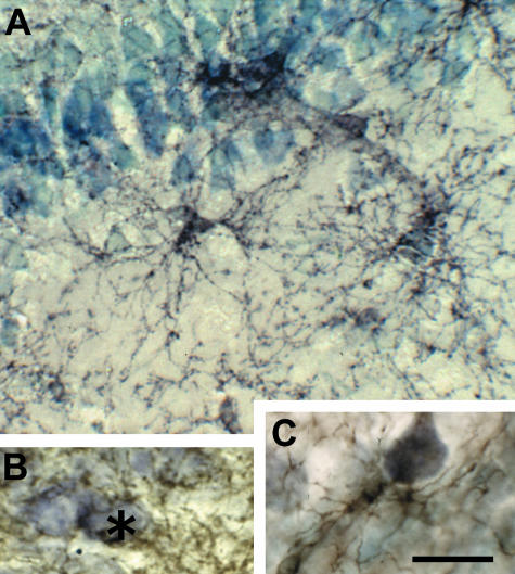
Fig. 2.
Synantocytes are closely associated with neurons. NG2-immunolabelled sections of adult rat brain counterstained with toluidine blue. (A) Synantocytes in the CA1 area of the hippocampus, some of which are directly apposed to pyramidal cell bodies, and extending processes through multiple layers. (B) Synantocyte processes forming a perineuronal network and enwrapping hippocampal neurons (asterisk). (C) Synantocyte apposed to and forming multiple contacts with a cortical pyramidal neuron. Scale bar = 25 µm.

