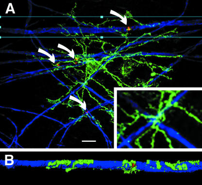
Fig. 4.
Synantocytes contact nodes of Ranvier. Confocal micrographs of whole-mounted anterior medullary velum triple immunofluoresecnce labelling for NG2 (green), myelin basic protein (blue) and ankyrin-3G (red). (A) Individual synantocytes contact multiple nodes of Ranvier (curved arrows) and their processes closely follow the path of myelinated axons (inset). All synantocytes observed in the velum formed similar associations with nodes of Ranvier. (B) Deconvolution shows the synantocyte process extending along the myelin sheath to form exquisite contacts with the paranodes and node of Ranvier. Scale bar = 10 µm in A, 30 µm in inset and 50 µm in B.

