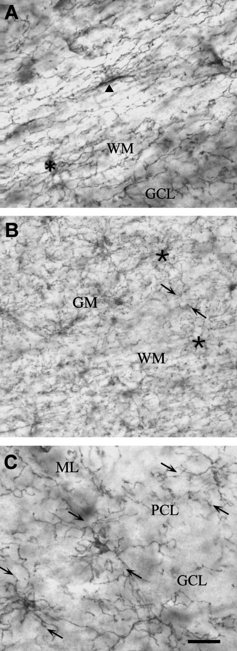
Fig. 5.
Synantocytes are interlaminar. NG2 immunolabelled sections of cerebellum (A, C) and cortex (B). (A) Individual cerebellar synantocytes with cell bodies in the white matter (WM, arrowhead) or at the interface between white and grey matter (GM, asterisk) extend processes into both. (B) Synantocytes in the cortical grey matter and subcortical white matter (asterisks) extend processes that traverse both layers and intermingle at the interface. (C) Interlaminar synantocytes extend processes into all layers of the cerebellum (arrows). Synantocytes are not specialized for either white or grey mater, and the same cells subserve both. Scale bar = 30 µm in A, C and 50 µm in B.

