Fig. 6.
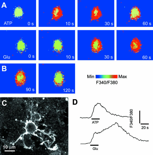
Synantocytes in vitro respond to glutamate and ATP with raised intracellular calcium. Explants of optic nerve glia were loaded with the calcium-sensitive dye fura-2 and imaged during bath application of ATP (A) or glutamate (B), and cells were identified at the end of the experiment by immunolabelling for NG2 (C). ATP and glutamate evoked a rapid increase in cytosolic [Ca2+]i in immunohistochemically identified synantocytes. (D) The response to ATP was transient and began to decay during exposure to the agonist, whereas the response to glutamate was slower to peak and was sustained after washout of the agonist.

