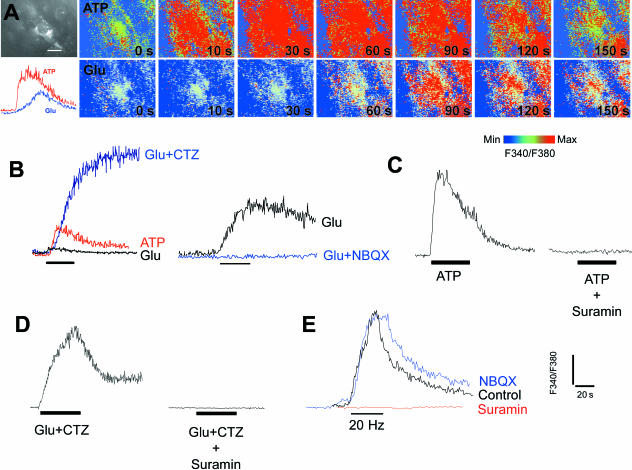
Fig. 7.
Calcium signalling in synantocytes in situ. Optic nerves were isolated intact and loaded with fura-2 for calcium imaging, and at the end of the experiment cells were identified by immunolabelling for NG2 (A). Both ATP and glutamate (Glu) evoked a rise in cytosolic [Ca2+]i in immunohistochemically identified synantocytes, although the response to ATP was rapid and transient, whereas that to glutamate was slow and sustained (A). These responses were analysed in greater detail in unidentified glia, but all cells in the optic nerve responded similarly (n > 100), and it reasonable to conclude that the findings reflect synantocytes as well as astrocytes and oligodendrocytes. (B) The response to glutamate was markedly increased by incubation with cyclothiazide (CTZ), which acts on AMPA glutamate receptors to maintain them in an open state, and was blocked by the AMPA receptor antagonists NBQX. (C) The ATP response was blocked by suramin, a general antagonist for P2X and P2Y purinoceptors. (D) Suramin also inhibited the response to Glu plus CTZ, indicating the increase in [Ca2+]i was partly mediated by ATP released in response to activation of AMPA receptors. (E) Electrical stimulation of the optic nerve at 20 Hz for 20 s induced an increase in glial [Ca2+]i that was inhibited by suramin but not by NBQX. The results indicate that glial calcium signals are evoked by ATP, presumably released from astrocytes in response to axonal electrical activity and activation of AMPA glutamate receptors, and support a primary role for ATP as a ‘gliotransmitter’ in the optic nerve.

