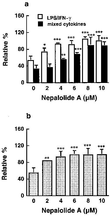
Figure 10.

The effect of nepalolide A on the degradation of IκB-α. C6 glioma cells were treated with 5 μg ml−1 LPS/100 units ml−1 IFN-γ (LPS/IFN-γ) or 5 ng ml−1 TNF-α/5 ng ml−1 IL-1β/100 units ml−1 IFN-γ (mixed cytokines) in the presence or absence of nepalolide A (2–10 μM) for 60 and 10 min, respectively (a). Astrocytes were treated with mixed cytokines plus or minus nepalolide A (2–10 μM) for 5 min (b). After treatment, cellular lysates were prepared and immunoblotting of intracellular IκB-α was performed. Results are means±s.d. (where large enough to be shown) from three independent experiments, and expressed relative to the cells at zero time. Significant differences between control and nepalolide A-treated cells are indicated by *P<0.05; **P<0.01; and ***P<0.001.
