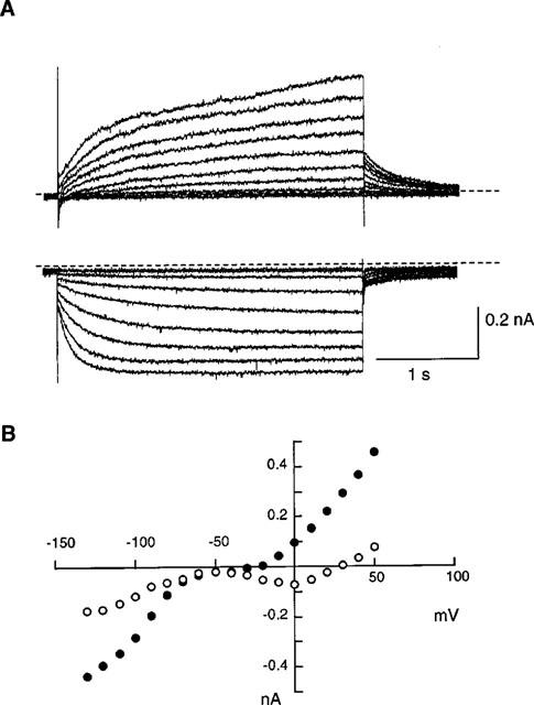
Figure 1.

Whole-cell currents of porcine sinoatrial node cells in the normal Tyrode solution. (A) Current traces recorded by applying 3 s depolarizing (upper traces) or hyperpolarizing (lower traces) pulses from a holding potential of −40 mV in 10 mV increments. Dotted lines indicate the zero current level. (B) Current-voltage relations for the initial currents (open circles) and the currents at the end of the 3 s pulses (solid circles).
