Figure 4.


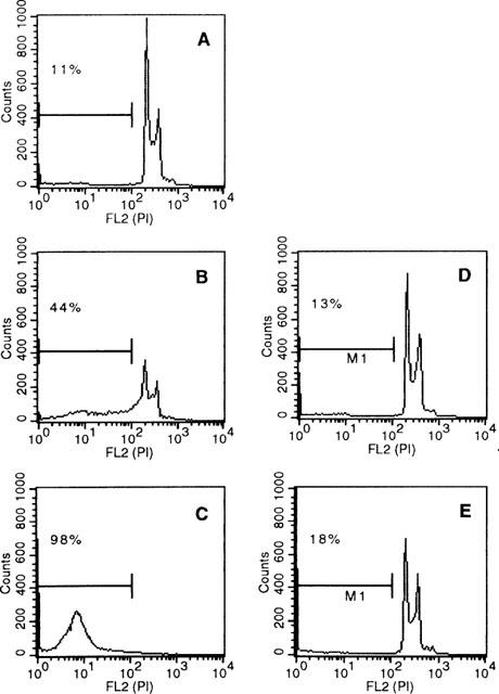
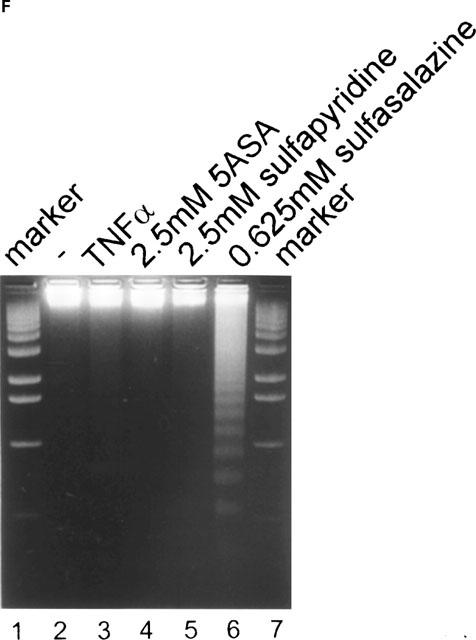
Sulfasalazine induces apoptosis in RBL5 T-lymphocytes as judged by DNA fragmentation. RBL5 cells were treated for 24 h with either (A) medium, (B) 0.625 mM sulfasalazine, (C) 2.5 mM sulfasalazine, (D) 2.5 mM 5ASA, or (E) 2.5 mM sulfapyridine. After ethanol fixation cells were stained with propidium iodide and analysed by FACS for the appearance of hypodiploid DNA. Numbers above the histogram markers indicate the percentage of apoptotic nuclei (broad hypidiploid peak) in a representative experiment (n=3). (F) Analysis of sulfasalazine induced DNA fragmentation pattern. RBL5 cells were treated with medium, 150 U ml−1 TNFα, 2.5 mM 5ASA, 2.5 mM sulfapyridine, or 0.625 mM sulfasalazine for 24 h. Genomic DNA was extracted and analysed on an ethidium bromide stained agarose gel. As molecular weight marker a 100 bp marker was used (Gibco). One representative experiment is shown (n=4).
