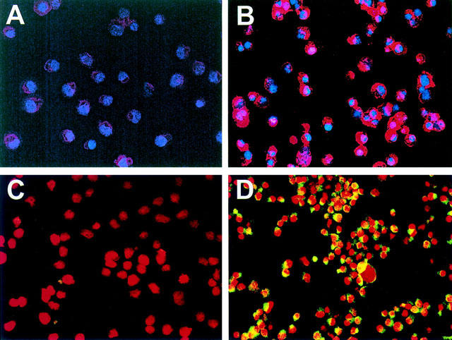
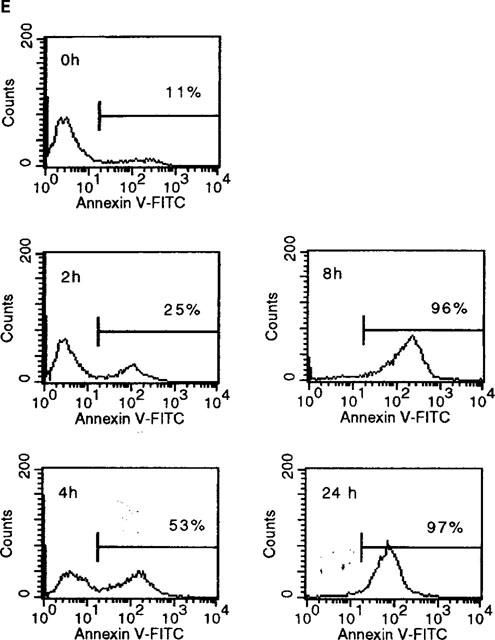
Figure 5.


Sulfasalazine induces apoptosis in RBL5 T-lymphocytes as judged by annexin V and Apo 2.7 staining. (A–D) Cells were incubated with medium (A,C) or 2.5 mM sulfasalazine (B,D) for 24 h. Cytospins were prepared and double immunofluorescence of annexin V binding (red) and Hoechst 33258 nuclear staining (blue) (A,B) or Apo 2.7 expression (yellow-green) and propidium iodide staining (red) (C,D) were performed. Fluorescence was observed and photographed with a fluorescence microscope (C. Zeiss, Oberkochen, Germany) equipped with epilumination. Magnification 400×. One representative experiment is shown (n=2). (E) For quantitative analysis of annexin V binding RBL5 cells were incubated with medium or 2.5 mM sulfasalazine for 2, 4, 8, and 24 h. Cells were stained with FITC-conjugated annexin V and analysed by FACS. 10,000 cells were analysed under each condition. Numbers above the histogram markers indicate the percentage of apoptotic cells in a representative experiment (n=3).
