
Figure 1.
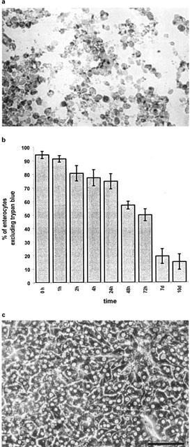
(a) shows a cytospin preparation of aspirated primary porcine enterocytes following 72 h in culture. Staining with Fast red shows polarized apical location of alkaline phosphatase of cells mutually attached with lateral surfaces. Proceeding culture time induces loss of cell polarity and circumferential staining for alkaline phosphatase. (b) records viability of duodenal enterocytes isolated according to protocol 5. Viable cells were found up to 10 days in culture. Viability was assessed by trypan blue exclusion. Data is shown as mean±s.d. of three dishes from three different isolations. (c) shows primary porcine sandwich hepatocytes at 96 h in culture by inverse light microscopy (bar=100 μm).