
Figure 4.
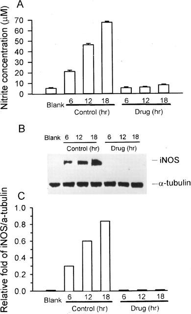
Western blot analysis of time-course protein expression of iNOS. RAW 264.7 macrophages were incubated in the presence of LPS (1 μg ml−1) plus IFN-γ (50 U ml−1) with or without 50 μM andrographolide for 6, 12, and 18 h, respectively. At the end of the incubation time, the culture medium was collected for nitrite assay (A) and extraction of total protein for iNOS protein and α-tubulin analysis (B). (C) Band intensities were quantified by densitometer. This experiment was repeated four times with similar results.