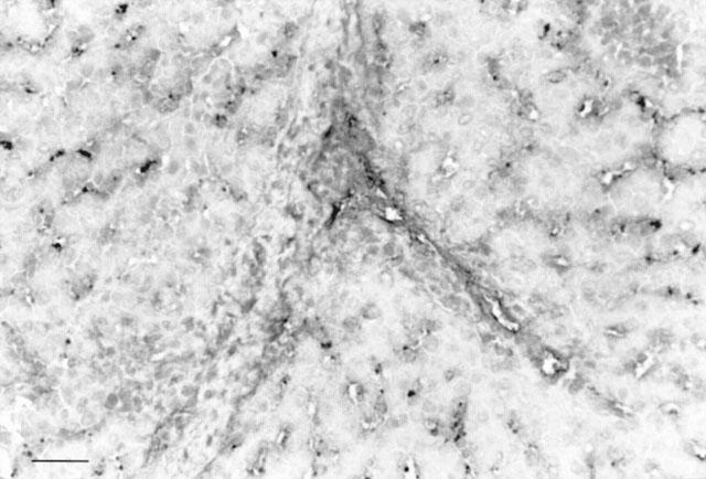
Figure 1.

Histological study. Minimal injury was constantly observed in the group perfused with xenogeneic serum. Only a few endothelial cells (<5%) of sinusoids and portal and central veins were stained after perfusion with trypan blue. Haematoxylin-eosin-safran staining. Bar=50 μm.
