
Figure 3.
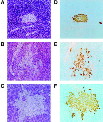
Histopathological analysis of pancreatic islets. Haematoxylin and eosin staining (A–C) and immunohistochemical staining by insulin antibody (D–F) were performed as described in Methods. Original magnification ×320: A, D, lean; B, E, ZDF control; C, F, ZDF treated with JTT-501 (147 mg kg−1 day−1).