
Figure 2.
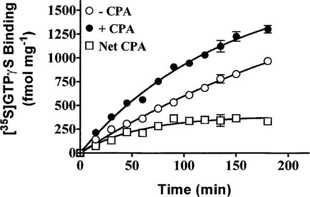
Time course of [35S]-GTPγS binding in the absence (−) and presence (+) of CPA. DDT cell membranes were incubated with 0.3 nM [35S]-GTPγS, 10 μM GDP and in the absence and presence of 1 μM CPA at 30°C for the times indicated. The net CPA stimulated-component of [35S]-GTPγS binding is the difference between the binding in the presence and absence of CPA. Each point on the graph is the mean±s.d. of triplicate determinations and is representative of three experiments.