
Figure 1.
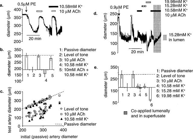
The ability of 10 μM ACh and 10.58 mM K+ to dilate rat mesenteric arteries. (a) A representative example of a pressurized artery that failed to dilate to on stepping from normal (5.88 mM) to high (10.58 mM) K+. 10 μM ACh was able to dilate this vessel close to its passive diameter both under control conditions (5.8 mM K+) and in high K+. (b) Mean data from seven similar experiments. (c) Scatter plot showing the relationship between the initial (passive) artery diameter (x-axis) and the level of tone of the arteries after PE constriction, in the presence of 10 μM ACh or high [K]o. In all arteries tested (n=30) ACh dilated to the passive diameter, whereas high [K+] only dilated to the passive diameter in nine arteries. The failure of high [K+] do dilate was not related to the diameter of the artery. (d) In three pressurized arteries where high (10.58 mM) K+ failed to produce dilatation, K+ was raised further in 4.7 mM increments by adding KCl from a 1 M stock. K+ failed to produce dilatation and at higher concentrations (19.98 mM) started to constrict. 10.58 mM K+ also failed to dilate when applied in the lumen. (e) Mean data (n=3).