
Figure 3.
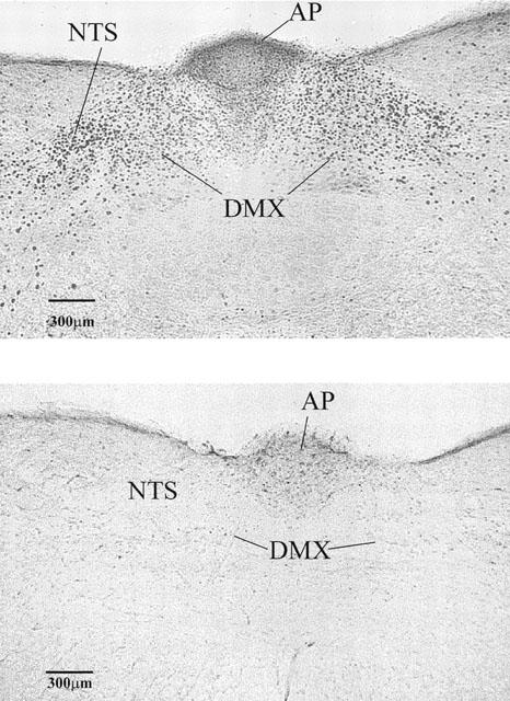
Photomicrographs of a representative transverse section of the caudal dorsal brainstem of Suncus showing fos-like immunoreactivity (dark stained cells) in one animal given RTX (100 μg kg−1 s.c. upper panel) and in another given its vehicle (lower panel). Note the intense fos-like immunoreactivity bilaterally in the nucleus tractus solitarius (NTS) and to a lesser extent in the body of area postrema (AP) in the RTX-treated animal. Fos-like immunoreactivity was noted in the dorsal motor vagal nucleus (DMX) but it was less dense than in the NTS.