
Figure 6.
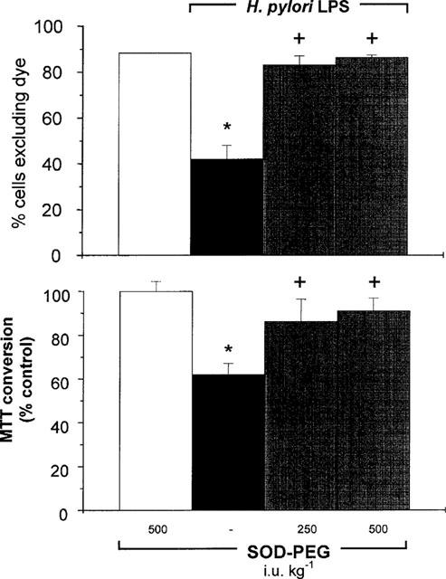
Percentage of viable isolated duodenal epithelial cells, as assessed by Trypan blue exclusion (per cent of cells excluding dye; upper graph), or MTT conversion (per cent control; lower graph), 5 h following challenge with H. pylori lipopolysaccharide (LPS; 3 mg kg−1, i.v.) in rat treated concurrently with saline or with superoxide dismutase conjugated with polyethylene glycol (SOD–PEG; 250–500 i.u. kg−1, i.v.). In control experiments, a group of rats received SOD–PEG (500 i.u. kg−1, i.v.). Data are mean±s.e.mean of 5–10 experiments, where * denotes a significant difference from the control (P<0.05) and + denotes a significant difference from the H. pylori LPS group alone (P<0.05).