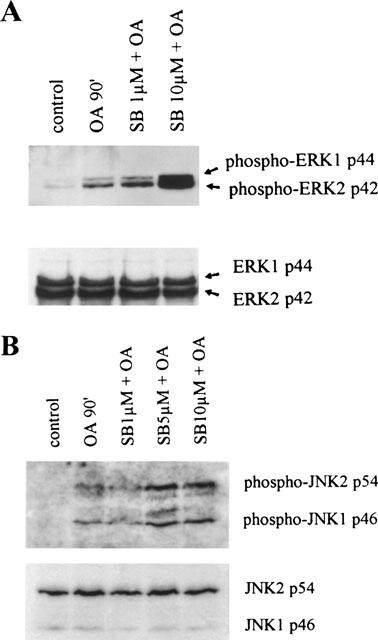
Figure 7.

SB203580 at 10 μM enhances phosphorylation of ERK1/2 and JNK1/2. TF-1 cells were cultured for 16 h in RPMI 1640 containing 0.1% FBS and subsequently stimulated with medium, OA (30 ng ml−1) for 90 min, or SB203580 (30 min pretreatment) in the indicated concentrations. Cell extracts were prepared as described in detail in ‘Methods'. Phosphorylated ERK1 and ERK2 (A) and phosphorylated JNK1 and JNK2 (B) are shown in the upper panels. Total ERK and JNK protein are shown in the lower panels and represent equal loading. Immunoblotting with anti-phospho-ERK/JNK and anti-ERK/JNK antibodies was performed by standard procedures and detection was performed using ECL immunodetection kit.
