
Figure 5.
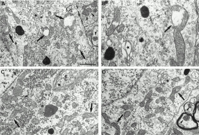
Electron microscopic photographs of lumbar spinal cord motoneurons from untreated pmn/pmn mice (A,B), control wild-type (C) and CGP 3466-treated pmn/pmn mice (100 nmol kg−1, 5 t w−1) (D). (A,B) In 42 day-old untreated pmn/pmn mice, a large number of mitochondria in the motoneurons displayed interrupted mitochondrial membranes (arrows) meanwhile outside the motoneurons they were well-preserved (arrowhead). Note the very large and swollen Golgi apparatus (star). (C) In age-mated healthy control wild-type mice, the mitochondria were well-preserved inside the motoneurons (arrow) as well in the other cells. Note the thin shape of the Golgi apparatus (star). (D) In 42-day-old pmn/pmn mice treated with CGP 3466B, most of the mitochondria were in excellent condition in the motoneuron cell body as well as in the dendritic structures. Despite the treatment with CGP 3466B the Golgi apparatus remained very swollen (star). Scale bar: 1 μm for A, C, D and 0.5 μm for B.