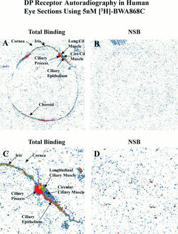
Figure 10.

Pseudo colour-coded images of DP prostanoid receptor binding sites in human donor eye sections using 5 nM [3H]-BWA868C. The respective magnifications for the various images shown in the panels, relative to the original images, were as follows: A and B = 3.4×; C and D = 8.5×.
