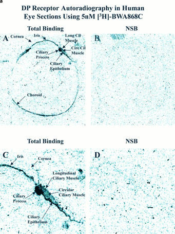
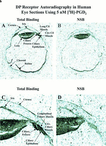
Figure 9.


Autoradiographic localization of DP-receptors in human eye sections using [3H]-BWA868C (a) and [3H]-PGD2 (b). (a) and (b) show total binding and non-specific binding profile in the whole eye sections at low power (top two panels; A,B) and at high power (bottom two panels; C,D; anterior segments of eyes) using 5 nM [3H]-BWA868C (a) and using 5 nM (3H)-PGD2 (b). The images are in black and white. The magnifications of the images relative to originals were: (a,b) panels A and B=4×; panels C and D=14.7×.
