
Figure 2.
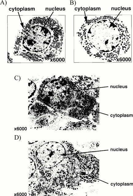
Electron micrographs (magnification ×6000) showing typical somatotrophs in (a,b) enzymatically dispersed pituitary cells and (c,d) the core region of a pituitary segment (diameter ∼1 mm) incubated for 3.5 h in the absence (a,c) or presence of annexin 1 ODN antisense (50 nM, b) or anti-annexin 1 mAb (diluted 1 : 15,000, d). Note (i) the electron-dense granules (diameter 200–350 nM) and extensive network of rough endoplasmic reticulum typical of somatotrophs, (ii) that in both preparations the cells appeared intact and well preserved at the end of the 3.5 h incubation and (iii) the appearance of the pituitary cells/tissue incubated with annexin 1 antisense ODN or anti-annexin mAb was indistinguishable from that of the controls; similarly, none of the other test substance tested (steroids, sense and scrambled ODNs or anti-spectrin α+β mAb) influenced the ultrastructure of the cells/tissue (data not shown).