
Figure 1.
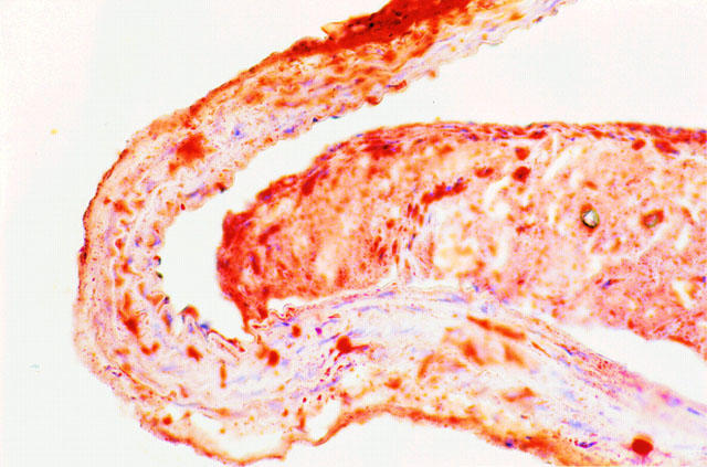
Transverse section of a representative thoracic aorta of an E°xLDLR° mouse after being fed a diet containing 21% fat and 0.15% cholesterol for 7 months, corresponding to the time of testing in vitro reactivity. The section is stained with Oil Red-O and counterstained with hematoxilin. Note the massive fibrofatty lesion obstructing a large part of the lumen. The plaque is partially detached from the vessel wall due to artefacts during sectioning and staining. Original magnification ×400.