
Figure 3.
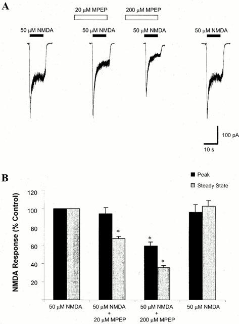
Effect of MPEP on NMDA-evoked whole cell current. (A) Representative traces of NMDA-evoked whole cell currents in the absence or presence of MPEP. Whole cell currents were recorded from cultured cortical neurones at 7–10 DIV, with voltage clamped at −60 mV in Mg2+ free solution. MPEP (20 or 200 μM) was delivered to the cell 30 s prior to delivery of 50 μM NMDA. (B) A summary of the effect of MPEP on NMDA-evoked current. Both peak and steady state responses were recorded. At a concentration of 20 μM, MPEP did not significantly alter peak NMDA current, but reduced the steady state current to 67.4±2.3% of control values (n=5, *P<0.05). At a concentration of 200 μM, MPEP significantly reduced both peak and steady state NMDA-evoked current to 59.4±4.2 and 35.1±2.5% of control values respectively (n=5, *P<0.05).