
Figure 6.
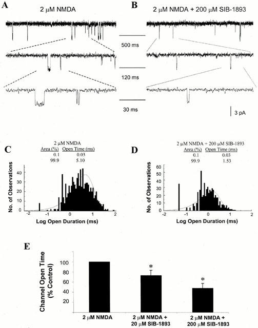
Effect of SIB-1893 on NMDA single channel open time. (A) Representative current traces evoked by 2 μM NMDA, obtained from an outside-out patch recorded from cultured cortical neurones at 7–10 DIV, with voltage clamped at −60 mV in Mg2+ free solution. Indicated channel current segments are shown on different time scale. (B) Representative current traces evoked by 2 μM NMDA and 200 μM SIB-1893 obtained from an outside-out patch. (C) Single channel open time was recorded from the patches in the presence of 2 μM NMDA and the distribution of the open intervals was fit with two exponentials with time constants and relative areas of 0.03 ms (0%) and 5.10 ms (100%). (D) Distribution of open intervals from the same patches in the presence of 2 μM NMDA and 200 μM SIB-1893. Distribution was fit with two exponentials with time constants and relative areas of 0.03 ms (0%) and 1.53 ms (100%). (E) Summary of the effect of 20 and 200 μM SIB-1893 on the NMDA-evoked single channel open time. In the presence of 20 μM SIB-1893, the channel open time was reduced to 73.02±10.02% of control values (n=5, *P<0.05). In the presence of 200 μM SIB-1893, the channel open time was reduced to 47.43±10.03% of control values (n=5, *P<0.05).