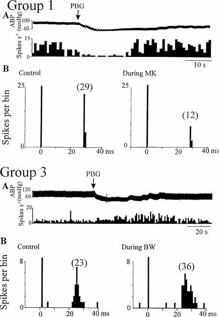
Figure 4.

Activity of NTS neurones activated by non-myelinated vagal nerve stimulation during cardio-pulmonary afferent stimulation and ionophoretic application of 5-HT2 receptor ligands. Group 1 cell: (A) Ratemeter record (1 s bins) showing the inhibitory effects of cardio-pulmonary afferent stimulation (PBG, arrows), (B) peri-stimulus time histograms (PSTH, 1 ms bins, 40 sweeps) on the same cell showing that application of MK-212 (30 nA, right) reduced the number of evoked spikes compared to pre-drug (left). Group 3 cell: (A) Ratemeter record (1 s bins) showing the excitatory effect of cardio-pulmonary afferent stimulation (PBG, arrows), (B) peri-stimulus time histograms (PSTH, 1 ms bins, 40 sweeps) of the same cell showing that application of BW-723C86 (40 nA, right) increased the number of evoked spikes compared to pre-drug (left). ABP: arterial blood pressure. Vertical bars at t=0 ms represent the stimulus artefacts and numbers in brackets are the number of evoked discharges counted.
