
Figure 3.
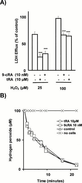
(A) Prevention by retinoids of H2O2-induced cytotoxicity. Confluent, quiescent CHMC cultured in 96-well microtiter plates, were incubated for 24 h with either 10 nM 9-cRA or 10 μM tRA. Cells were then washed and incubated for 24 h with either 25 or 100 μM H2O2. Control cells were incubated in parallel but without adding retinoids and H2O2. Cytotoxicity was quantified by measuring LDH efflux from the cytosol of the damaged cells to the supernatant. Results were expressed as the per cent of the control value (LDH released by control cells incubated with 2% Triton-X-100 was considered as 100% LDH efflux) and are mean±s.d. of three separate experiments in quadruplicate. As shown, 10 nM 9-cRA and 10 μM tRA prevented in a similar extension the H2O2-induced loss of cell viability (***P<0.001). No differences in LDH release were found beween control cells and cells incubated with 9-cRA or tRA (results are not shown in the figure). (B) Consumption of H2O2 by cells incubated with retinoids. Confluent, quiescent CHMC cultured in 35 mm Petri dishes, were incubated for 48 h with either 10 nM 9-cRA or 10 μM tRA in order to stimulate maximally antioxidant defences. Cells were then washed and incubated with 100 μM H2O2 from 0 to 22 min. Control cells were incubated in parallel but without adding retinoids. At the times shown in the figure, H2O2 was measured in an aliquot of medium by the horseradish peroxidase-mediated oxidation of phenol red method. Results are the mean±s.d. of three separate experiments in triplicate. No statistically significant differences were found in the consumption of H2O2 between the three experimental groups (ANOVA P>0.05).