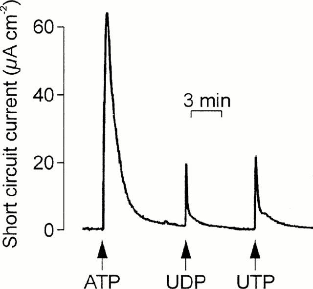
Figure 2.

Evidence for multiple receptor subtypes in the apical membrane. ISC was recorded whilst cells were exposed to apical ATP followed by UDP and then UTP. Arrows denote the addition of the appropriate nucleotides (100 μM); these were present throughout the remainder of the experiment. Essentially identical records were obtained in four instances.
