Figure 5.

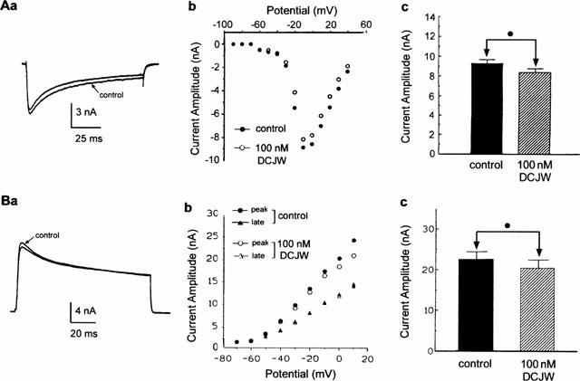
Absence of blocking effect of DCJW on inward calcium and outward potassium currents. Whole-cell high-voltage activated (HVA) calcium current (Aa) elicited by 100-ms depolarizing voltage pulse to −10 mV from a holding potential of −100 mV, before (control) and after application of 100 nM DCJW. (b) Current-voltage relationships constructed from values of peak current plotted as a function of test potentials, in control and after application of 100 nM DCJW. (c) Histogram comparing the peak HVA current recorded before (control) and after bath application of DCJW (n=4). (Ba) Superimposed global outward potassium current traces recorded in saline containing 100 nM TTX (control) and following application of 100 nM DCJW. Outward currents were evoked by depolarization to +10 mV (100 ms in duration) from a holding potential of −80 mV. (b) Current-voltage relationships of both peak and late outward potassium current before and after application of 100 nM DCJW. (c) Histogram comparing the effect of 100 nM DCJW on the peak outward current measured at +10 mV. In both cases, the Student's t-test (•) was used to indicate that the difference was not significant (P>0.05, n=4).
