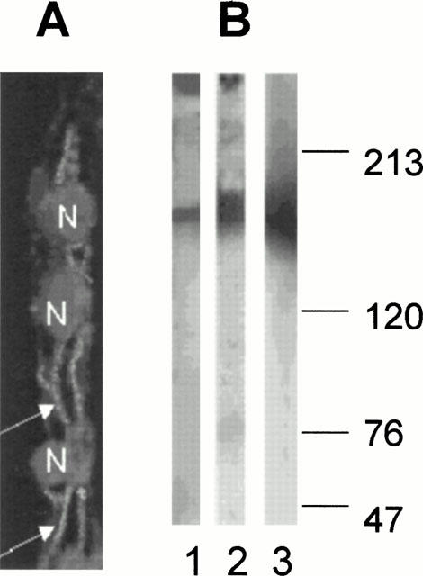
Figure 2.

(A) Confocal image of porcine brain capillaries double stained with the monoclonal antibody versus Pgp and propidium iodide for nucleus (N) staining. Pgp staining can predominantly be observed at the lumenal membrane of the brain capillary endothelial cells (arrows). (B) Western blot detection of Pgp in isolated intact capillaries (1) and 7-day-old cultures of brain endothelial cells (2). Pgp-positive control (MDR+)-P388 cells (3). The numbers indicate molecular weight markers (in kD).
