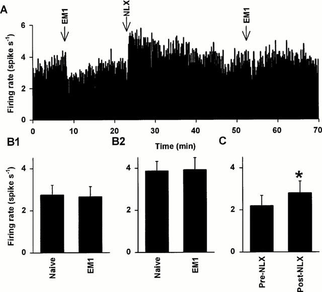
Figure 3.

Effects of chronic i.c.v. endomorphin 1 on the activity of oxytocin and vasopressin cells in urethane-anaesthetized rats. (A) The firing rate (averaged in 10 s bins) of a SON oxytocin cell (excited after 20 μg kg−1 CCK i.v.; not shown) recorded from a urethane-anaesthetized rat infused with i.c.v. endomorphin 1 for 5 days. This cell was inhibited by acute injection of endomorphin 1 (100 pmol, i.c.v.), but to a lesser extent than in endomorphin 1 naïve cells (Figure 2A,D). Similarly to its effects on endomorphin 1 naïve cells, naloxone (5 mg kg−1, i.v.) caused only a small increase (relative to pre-EM1) in the firing rate of the cell, and markedly reduced its responsiveness to a second injection of the same dose of endomorphin 1. (B) The spontaneous firing rates (+s.e.mean) of SON oxytocin (B1) and vasopressin (B2) cells recorded from urethane-anaesthetized endomorphin 1 naïve (oxytocin: n=17; vasopressin: n=25) and endomorphin 1 pre-treated (EM1; oxytocin: n=14; vasopressin: n=15) rats. There were no significant differences in the initial firing rates of oxytocin cells, or of vasopressin cells, between endomorphin 1 naïve and endomorphin 1 pre-treated rats, indicating that both cell types were tolerant to the inhibitory effects of i.c.v. endomorphin 1. (C) The mean firing rate of SON oxytocin cells recorded from endomorphin 1 pre-treated rats (n=9) 5 min before and after naloxone injection (NLX, 5 mg kg−1, i.v.), showing a small increase in firing rate after naloxone (*P<0.05; paired t-test).
