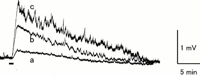
Figure 4.

Ventral root depolarization evoked by maxadilan. Potential was recorded extracellularly from the L4 ventral root of an isolated spinal cord from a 1-day-old rat. Maxadilan was applied to the medium superfusing the spinal cord during the period (60 s) indicated by the bar. (a) 0.01 μM (b) 0.1 μM (c) 1 μM.
