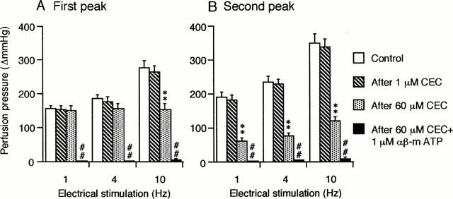
Figure 3.

Effects of CEC and αβ-m ATP on the first (A) and the second peak (B) of the vasoconstrictor responses to periarterial electrical nerve stimulation in the canine splenic arteries. The vessels were electrically stimulated by 30 s trains of pulses at 10 V amplitude and 1 ms pulse duration, with a frequency of 1, 4 or 10 Hz. Data are presented as mean±s.e.mean (n=12). **P<0.01 as compared with the control group. ## P<0.01 as compared with the preceding group.
