
Figure 8.
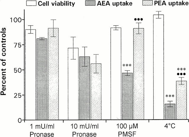
The effects of treatment with Pronase, PMSF and 4°C upon cell viability, AEA and PEA transport in Neuro-2a cells. The cells were incubated with the indicated concentration of Pronase or PMSF (b), followed by the addition of 10 μM [3H]-AEA or 20 μM [3H]-PEA. After 15 min at 37°C or 4°C, the cellular uptake of AEA and PEA and the cellular release of LDH or cellular MTT reduction were determined. Data are plotted as percentage of untreated control cultures and represent means±s.e.mean (n=3 – 8). Statistically significant differences (one-way ANOVA with Bonferroni multiple comparison test) are indicated: ***P<0.001 when cellular AEA or PEA uptake is compared with corresponding cell viability values, and •••P<0.001 between AEA and PEA uptake at respective treatment.