Figure 1.

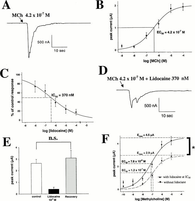
(A) Sample trace of a Ca-activated Cl current (ICl(Ca)) induced by 10 s administration of MCh at approximately EC50 (4.2×10⁻⁷ M) in an oocyte expressing the muscarinic m3 receptor. Peak current is 1.12 μA. (B) MCh evokes ICl(Ca) in a concentration-dependent manner. EC50 is 4.2±0.4×10⁻⁷ M, Emax is 2.0±0.4 μA. (C) Lidocaine inhibits MCh (at EC50)-induced ICl(Ca) in a concentration-dependent manner. IC50 is 3.7±0.8×10⁻⁷ M. (D) Sample trace of a m3 response elicited by stimulation with MCh (at EC50) in the presence of lidocaine at IC50. Peak current is 0.48 μA (A). (E) Mean±s.d. of m3 responses elicited with MCh (at EC50). Left bar indicates the control response, middle bar represents the response after 10 min incubation in 10−4 M lidocaine, and right bar represents the response after a 10 min incubation in 10−4 M lidocaine and another 10 min wash with Tyrode's solution. The third response is not significantly different in size from the first one, indicating reversibility of lidocaine inhibition. (F) Concentration-response curve for MCh on m3 receptors in the presence or absence of lidocaine (at IC50). Lidocaine inhibition cannot be overcome with maximal agonist concentrations (Emax remains inhibited by 36%) suggesting a primarily non-competitive antagonism.
