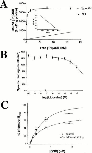
Figure 2.

(A) Characterization of [3H]-QNB binding to membranes prepared from CHO cells, stably transfected with the rat muscarinic m3 receptor. The saturation curve and Scatchard analysis conform to a single site model with a Kd of 0.15±0.03 nM (n=5) and Bmax of 3517±154 fmol mg−1 protein (n=5). (B) Effects of lidocaine on specific binding of [3H]-QNB to muscarinic m3 receptors. IC50 is 78±9 mM (n=5). Lidocaine at functionally determined IC50 (3.7×10⁻⁷ M) does not affect agonist binding. (C) Binding curves for [3H]-QNB to m3 receptors in the absence (control, – • – ) and presence (–□amp;–) of lidocaine at IC20 for binding effect (5×10⁻⁴ M). Whereas Kd is not significantly shifted (0.57±0.01 nM in the absence and 0.48±0.04 nM in the presence of lidocaine), Bmax decreases in the presence of lidocaine by 33.5±3.8% of control, from 1482.1±88 to 984.3±45.5 fmol mg−1 protein.
