
Figure 3.
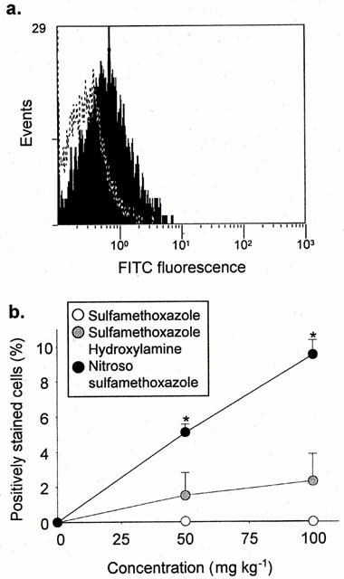
In vivo cell surface haptenation of rat PBMC after administration of sulphamethoxazole and its metabolites for 1 h. PBMC were isolated and haptenation was determination by flow cytometry using a specific anti-sulphamethoxazole antibody. The majority of cells were gated for analysis and the forward threshold was raised to exclude debris. (a) Flow cytometric traces showing differential staining of PBMC populations from animals administered DMSO (dotted line) and nitroso sulphamethoxazole (shaded; 100 mg kg−1). (b) Graph showing cell surface haptenation of rat PBMC after administration of nitroso sulphamethoxazole, but not sulphamethoxazole hydroxylamine or sulphamethoxazole. The results are presented as the percentage of positively stained cells from four rats, incubations carried out in triplicate. Statistical analysis compares drug-treated rats with animals administered DMSO (*P<0.05).