
Figure 2.
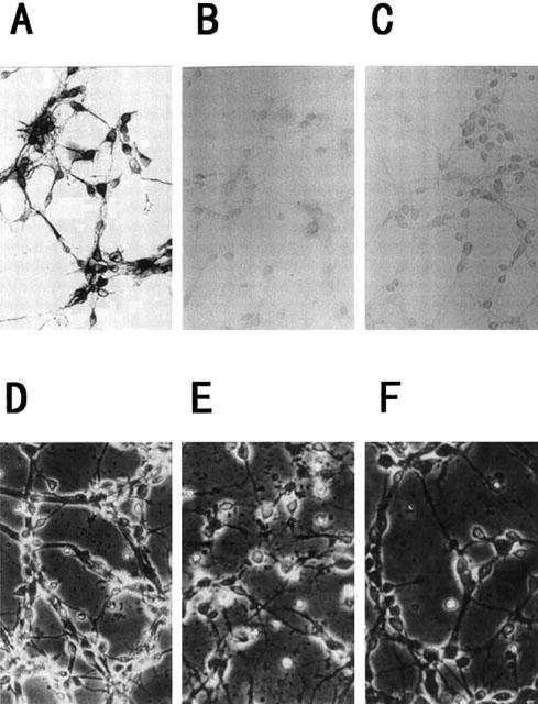
Morphologic changes in cortical neurons by Aβ(25 – 35). Immunocytochemical analysis for anti-MAP2 (A), anti-GFAP (B) or OX-42 (C) was performed on the present cortical cultures. Magnification, ×95. Cortical neurons were treated with vehicle control (D), 10 μM Aβ(25 – 35) (E), or 10 μM Aβ(25 – 35)+10 μM S-2474 (F). Neurons were examined by phase-contrast microscopy 48 h later. Magnification, ×95. Vehicle control is treated with culture medium containing 1% deionized water and 0.1% DMSO. Aβ control is treated with culture medium containing 10 μM Aβ (25 – 35) and 0.1% DMSO.