
Figure 5.
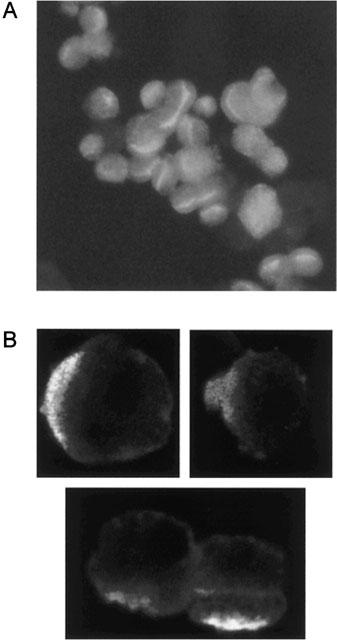
(A) A representative microscopic image of rat kidney PTC after staining with an antibody against alkaline phosphatase and a fluorescent secondary antibody. Staining is strongest along the luminal membrane of the PTC, confirming the maintenance of cell polarity. (B) Immunolocalization of Mrp2 in rat kidney PTC. Cells were immunostained with polyclonal antibodies against Mrp2 and a fluorescent secondary antibody. Mrp2-specific immunofluorescence is observed at the luminal membrane, confirming the apical localization of the transporter.