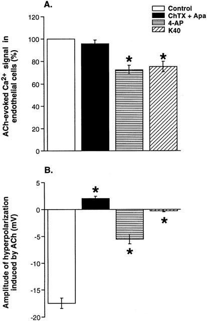
Figure 3.

Comparison of the effects of K+ channel blockers on acetylcholine-evoked changes in endothelial cells Ca2+ signal (A) and SMCs membrane potential (B). Responses to ACh (1 μM) were measured in the absence (control) and in the presence of charybdotoxin and apamin (ChTX+Apa, 0.1 μM), 4-aminopyridine (4-AP, 5 mM) or in physiological solution containing 40 mM of KCl (K40). Endothelial cells Ca2+ signal was expressed as a percentage of the maximum amplitude of acetylcholine-evoked responses in the absence of test drugs. Data are presented as means±s.e.mean. Asterisks denote a statistically significant difference from control values (P<0.05).
