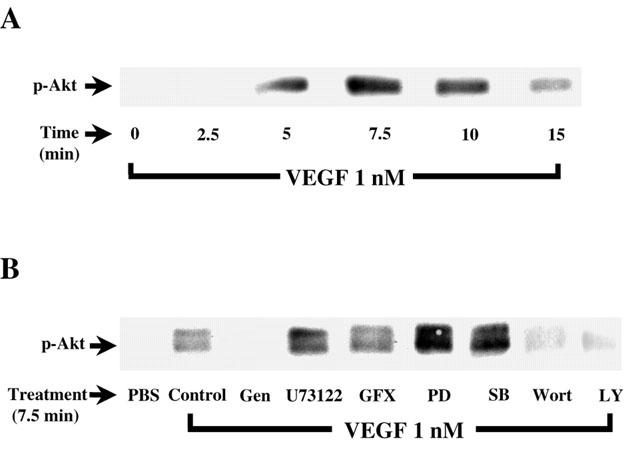
Figure 7.

Effect of VEGF on PI3K pathway activation. (A) Confluent BAEC were stimulated with VEGF (1 nM) for various periods of time. Cells were lysed and separated on a 10% SDS – PAGE. Western blot analysis was performed with an anti-phospho-Akt antibody. (B) Western blot determination of the phosphorylated form of Akt reveals the effect of VEGF-induced Akt phosphorylation. Experiments were carried out as in Figure 6A, except that BAEC were pretreated 5 min with genistein (Gen, 10 μM), U73122 (10 μM), GF109203X (GFX, 1 μM), PD98059 (PD, 10 μM), SB203580 (SB, 10 μM), Wortmannin (Wort, 100 nM) or LY294002 (10 μM) prior to a 7.5 min treatment with VEGF (1 nM).
